Jumat, 09 Agustus 2013

Brain development; neurogenesis, apoptosis and synaptogenesis

I do not know you but  I find incredible the way that nature uses the energy and resources to create masterpieces, as flowers, honey bees or brains.

An example of this is brain development, since the nervous tissue formation begins with the formation of a simple tube, called neural tube from the neuroectoderma induction (this is part of the ectoderm that is the outermost cell primary embryo that originates the central and peripheral, nervous systems including some glial cells), this process occurs as part of human neurodevelopment between the third and fourth gestational week and wrapped the most fascinating functions that it is possible to imagine.

But the road is not simple, since once formed the neural tube occurs a differentiation in three dimensions: the first leads to the spinal cord, the second will give rise to stem and brain stem and the cerebellum, while the third portion will develop the cerebral hemispheres.

This stage is called a proencephalic process that occurs between the fifth and tenth gestational week and during which an active neurogenesis (neuron development) and this develops from the neural precursor cells, which have a special feature and is not mature and do not proliferate, because it will have to wait for the next moment for such differentiation (Poch, 2001).

Between the eighth and tenth eighth gestational week, occurs an active neuronal proliferation, the precursor cells begin to differentiate to produce new precursor cells and neuronal cells different of neurons and glial cells (astrocytes and oligodendrocytes).

The speed of proliferation in this period that occurs between the 2nd and 4th month of intrauterine life is impressive since they are around 200,000 neurons per minute, as fast as any kind of thought, this cell proliferation, is known as neurogenesis, this process occurs slowly, and after going through several cycles of cell division, this is stopped, since as we know, that in excess is bad.

Even if it is unknown what starts and then stops the mechanism of proliferation in any region, it is clear that the periods are rigidly determined, although we know it depends on neural factors specific to the region of the brain where it occurs and functions to be exercised, by between what 3rd and 5th month occurs the migration of neurons guided by processes glial based on chemical signals and neural growth factors, mediated by regulatory genes that determine the activity of other genes in a defined sequence and for precise periods and in specific regions.

What has already been observed is that differentiated cells begin to emigrate from ventricular areas (Central) to the more peripheral areas of the brain (neocortex) development.

This is a specific order, which begin first occupy the deeper layers of the cortex layers, while those that start later, occupy the uppermost layers.

This radial migration of neurons to the periphery, uses glial cells as a guide since these form a scaffolding that facilitates the movement of neurons. Neuronal migration occurs mainly in two regions in the thalamus and hypothalamus, where the oldest neurons are pushed by more new neurons, by which the first will be located in the periphery. On the other hand, in regions of the brain structure of laminar, as it is the case of the cortex and the cerebellum, neurons more young people migrate to break through to the oldest, whereupon the latter will sit closer of the neuroepithelium and the more young people on the periphery.

Neuronal migration process takes place between the 10th second and the twenty fourth gestational week, but it is noteworthy that not all cells survive since during neurogenesis and neuronal migration, approximately 50% of neurons undergo apoptosis, meaning that cells die in a programmed way, probably because they do not follow the correct course of emigration or because they do not receive the stimuli of the rest of the networks that are created, but the correct answer is still a mystery.

A certain proportion of the neurons that survive (20%) Trek horizontally and one after radial migration, to allow the formation of lamination (segmentation) cortex, it is so neurons looking his way, motivated by chemical stimuli (neurotrophic factors), extending its structure in one of its ends, resulting in the so-called axonal growth cones.

Simultaneously with the neuronal migration occurs in synaptogenesis (formation of synapses), although this is much more intense between the twelfth and the twelfth fourth gestational week, but persists in a very active way until the eighth or ninth month postnatal (Avaria, 2005; Sanhueza, Nieto and Valenzuela, 2004, Sagan, 2003; Poch, 2001).

It is interesting to note that prenatal synaptogenesis is mainly determined by the genetic heritage of the individual. However, in the stage postnatal synaptogenesis is also affected by sensory experiences, particularly by the learning process and the stimulation from the environment.

Neurogenesis and the subsequent stages associated with this process morphogenic lead to the formation of approximately 100 billion neurons in the adult brain and several trillions of synapses. This implies that a significant number of the 30,000 genes that we have must be involved in this complex process, expressing together in simultaneous or sequential form.

However, she has still not been achieved understand this prodigious process, because a region possessing 20,000 genes, is only 302 neurons and nerve tissue that form is far from having the functionality of the human brain (Sanhueza, Nieto, and Valenzuela, 2004).

So as a result of the differential expression of genes in the course of time, and in different cells of the embryonic structures, given the development process, but this depends on much more than a base sequence of the genome and the history and composition of the genome of the parents.

While the genome gives specificity and individuality that new body, its expression depends not only on bases of DNA, but also the protein and environmental environment in which it is (Vasquez Laslop and Velázquez Arellano, 2004).

As son as intrauterine development finish, we see the birth of a new human being, but even it is true that the neural cells are there, ready to connect with each other, will take between 2 and 3 years until they manage to do it logically, because the relationship between them depend on the needs of the environment.

When brains of the babies are ready to see the light,  they work but they are very immature, partly because the brain needs lot of flexibility to adapt and reply to the environment. This prevents them from performing tasks that other mammals in a few days or weeks after birth, for example, human babies require much effort to set the look, or control the trunk to sit. However, are born with reflexes that will enable them to adapt to the new environment, as a reflection of suction, that will give you food that requires to help its development, although there are others like blinking and crying that will provide you the opportunity to have contact with caregivers (Quintero Gallego, Manaut, Rodríguez, Pérez - Santamaria and Gómez, 2003) .

The number of cells in the fetal brain is between 30 and 70% higher than the number of neurons in the adult. Surplus cells survive for a period of days to weeks, after which, on its own, starts a cascade of degenerative changes and a physiological process of programmed cell death. In the picture below, it is possible to observe the differences between birth and two years of development, although it seems that increased neuronal tangle, in reality there are what are they are less neurons with larger number of neural networks, connections between neurons, i.e. interneuronal communication, which allows a more robust network that ensure more specific skills.

In this sense, found that the selective removal of the synaptic connections, is a fundamental process in the cognitive development of the child, as has been observed relationship between changes in the gray matter of the frontal lobe and the evolution in the performance of cognitive tasks (Chapel, Romero, Maestu, field, Fernandez, Gonzalez Marquis, Fernandez and Ortiz, 2004;) Hubel and Wiesel, 1970).

At this point, important thing is to start the race to connect to neurons. Although the neuronal pruning will be inevitable, since this is a process of survival of the fittest under the Darwinian principle that everything that is not used is discarded, neurons that fail to create medium-term goals that enable adaptation, they will be destroyed, the important thing for the brain is to achieve the process critical enabling that new creature to make it into the world. The critical process will be listening, see (to later make discrimination environment and information) stand up, little by little to control their own weight (around the year of born), recognize the natural mother language  for later use it to communicate and finally walk.

Some say that the first two years are the most important for the education of children, and there are those who insist that math, music or foreign languages are important in this critical period of development, however, there is no evidence that this is vital for the process of cognitive development, because they can be perfectly learned once the most important process occur.

What is achieved with this is process of brain, organization which begins six months of gestation and lasts throughout life. Although the rhythms vary, thus, as already mentioned, during the last trimester of pregnancy and the first two years of life, the pace of organization is quickened, then becomes less rapidly to 10 years of life, to continue so slow during the rest of the life.

During the acceleration phase, occurs a large increase of dendritic extensions and small branch, which has been called dendritic arborization, that form numerous synapses, so that all cells and its extensions are arranged in layers and orient themselves, at the same time causing programmed cell death and differentiation and specialization neuronal, this depending on the interactions with the environment and genetic factors.

So crests of the neuronal branches are, density peaks occur at different ages, but also in different brain areas. Thus one fast and dense development both in the visual cortex and the hearing between the 3 and 4 postnatal months and maximum density, around the year of life can be observed. Against, the growth of the prefrontal area is presented at the same age, but the peak is reached until after the first year of life.

The only exceptions are the granulated cells of the olfactory bulb, cerebellum and hippocampus, which continue its genesis after birth and continue for life (León Carreón, 2003).
However, there is also a regressive phase, which presents a selective loss of synapses, this occurs after periods of great intensity. In the same way that the synaptic formation crests, decreases the time of reduction varies between different regions, for example, the visual cortex synaptic density, between 2 and 4 years, while the prefrontal cortex, takes place between 10 and 20 years of age approximately (Poch, 2001).

These periods coincide with the stages of cognitive development widely explained by Jean Piaget, if the stages of brain development, equate either dendritic arborization of the gray matter development, coincide with the periods marked for cognitive progress. From both positions, the environment will play a leading role, but in the case of brain development will be added, intrauterine infections, chromosome pathologies or nutritional deficits that may directly affect the development within the mother's uterus.

Myelination, (which is an overlay of the neural connections by a specialized membrane that allows a proper transmission of nerve impulses), is essentially a post natal process, occurring in cycles, with a default orderly sequence that contributes greatly to improve the functionality of the brain because it produces an increase in the speed of nerve impulse conduction.

In this sense has been found that there is an increase in white matter during childhood, which probably reflects the increase in myelination (Quintero Gallego, Manaut, Rodríguez, Pérez - Gómez and Santamaria, 2003;) Chapel, Romero, Maestu, field, Fernandez, Gonzalez Marquez, Fernandez and Ortiz, 2004; Avaria, 2005).

However, this process, as well as the maturation process, occur at different times, in particular found that the projection areas, mature before the associative, so the latest in purchasing a myelinated appearance are frontal parietal and occipital lobes (Quintero Gallego, Manaut, Rodríguez, Pérez - Gómez and Santamaria, 2003; Chapel, Romero, Maestu, Campos, Fernandez, Gonzalez Marquis, Fernandez and Ortiz, 2004; Avaria, 2005).

References:

Avaria, M. A. (2005)  Aspectos biológicos del desarrollo psicomotor.  Revista de Pediatría. Electrónica. 2 (1). Disponible en red: http://www.revistapediatria.cl/vol2num1/pdf/6_dsm.pdf

Capilla, A.,  Romero, D.,  Maestu, F., Campo, P., Fernández, S., González Márques, J., Fernández, A. y Ortiz, T. (2004) Emergencia y desarrollo cerebral de las funciones ejecutivas. Acta. Esp. Psiquiatr. 32 (2) 377 – 386.

Hubel D., Wiesel T. (1970) The period of susceptibility to the physiological effects of unilateral eye closure in kittens. J Physiol. 30 (4) 206- 212.

León Carrión, J. (2003) Células madre, genética y neuropsicología. Revista Española de Neuropsicología. 5 (1) 1-13. 

Poch, M.L. (2001) Neurobiología del desarrollo temprano. Contextos educativos. 4. 79-94.

Quintero Gallego, E., Manaut, E. , Rodriguez, E., Pérez – Santamaría, J.,  y Gómez, C. (2003) Desarrollo diferencial del cuerpo calloso en relación con el hemisferio cerebral. Revista Española de Neuropsicología. 5 (1) 49-64.

Sanhueza, J., Nieto, S. y Valenzuela, A.  (2004)  Acido docosahexaenoico (dha), desarrollo cerebral, memoria y aprendizaje: la importancia de la suplementación perinatal. Rev Chilena Nutrición. 31 (2) Disponible en: http://dx.doi.org/10.4067/S0717-75182004000200002 

Sagan, C. (2003) Los dragones del Edén: especulaciones sobre la evolución de la inteligencia humana. Barcelona. Crítica.

Stromswold, K. (1995) The cognitive and neural bases of language acquisition: The cognitive neurosciences. Cambridge, MA: MIT Press.

Vásquez  Laslop, M. y Velázquez Arellano, A. (2004) Genómica y el desarrollo de un nuevo individuo. En A. Velazquez (2004) Lo que somos y el genoma humano: des-velando nuestra identidad. Ediciones científicas universitarias. UNAM. FCE.

Kamis, 18 Juli 2013

Genes and genetic disorders

If we think about how amazing it has been  the Human Genome project, how much we have learned from it and everything that has been generated so far, it's impossible to ignore the value of genomics, for example,  initially was thought that our 46 chromosomes were composed of 100,000 protein-coding genes, enzymes, hormones and overall regulatory sources of all life processes that shape human characteristics but now is possible to begin to manipulate all of them  to heal syndromes and diseases.

Chromosome 22 was the first to be fully identified 545 genes having assets, followed later by genetic mapping or sequencing of chromosome 21, with only 225 active genes, which opened the possibilities for genetic treatments medical situations as Down Syndrome, a form of Alzheimer's disease and various cancers.


Thus began the race to find a specific gene for each of the major conditions that afflict humans. At the same time, it have been found the relationship between the production of proteins and some genes that create on protein production and others that inhibit the production of the same.


In this sense it was found some genes associated with autism spectrum syndromes such as 15q11-q13, 2q37.3 or UBE3A for Angelman syndrome, which has also been associated with the GABRB3 gene, whereas for the syndrome Williams is the gene 7q11.23; MeCP2 for Rett syndrome, to mention only the best known developmental syndromes.


However, it is known that there are more syndromes, such as West syndrome that is associated with the ARX gene, or even lower prevalence may be mentioned De Lange syndrome or Crohn's disease, whose genetic alteration is in 5p13.1 gene.


Talking about less common syndromes we can talk about Smith-Magenis syndrome which is associated with  17p11.2 gene, or the VCFS 22q11 gene related, with myotonic dystrophy which is a multisystem disease whose genetic alteration is associated with the repetition CTG trinucleotide on chromosome 19.


While the tuberous sclerosis complex (TSC), which is an autosomal dominant inherited disease which may be due to the mutation of two distinct genes: TSC1 (9q34) and TSC2 (16p13.3).


Timothy syndrome caused by a mutation in the gene CACNA1C located in 12p13.3, other forms of alteration as 10p terminal deletion, has been associated with a phenotype similar to DiGeorg syndrome causing hiccups hyperparathyroidism, deafness sensory abnormalities stones.


Moreover, it has been  studied the gene involved with 45X/46XY mosaicism, or we can mention Cowden syndrome is associated with PTEN gene, which is located at 10q23.3.


Another group of genetic disorders like Goldenhar syndrome are related  to deletion 5q (Artigas-Pallares, Gabau-Vila & Guitart-Feliubadaló, 2005), all associated with developmental disorders.


Whereas if you look at the studies on cancer have been found, for example for brain cancer, NFkB protein with much higher activity than in normal brains or HER2 protein in lung and breast cancer, BCR- AB1 for chronic myelogenous leukemia, RAS, for several types of cancer, B-RAF for skin cancer, BCL-3 for lymphoma; RB1 for retinoblastoma, HNPCC for colon cancer and endometrial, the p53 gene causes cell suicide and is associated with cancer of the lung, colon, breast and brain (Collins and Barker, 2008).


It's not possible to forget disorders of cognitive processes such as Alzheimer's,  so it's possible to find genetic associations with cognitive processes, although further studies are not conclusive, for instance mentioned the IGF2R gene associated with intelligence, which is a very popular topic.


That is why around 1980 Dr. Plomin, from Institute of Psychiatry, at London, England, began his research on genes and intelligence relationship, since observed that two people with the same genes correlate as much as the same person performing an intelligence test with a year of difference, so he realized that identical twins who live apart are very similar or identical in intelligence tests that identical twins who live together, which is why it is considered that the environment influences the development of children, however it has become clear that genes can shape the brain in ways that make individuals better or worse to answer an intelligence test.


Despite this, Plomin suspect that more markers are needed to find the genes for intelligence, but states that exist many difficult to replicate details to affirm conclusively that have these genes (Silverman, 2008).


More consistently it was found the  called
language gene, which from the genetic standpoint is associated with the specific disorder of the cognitive process, which seems to be due to alteration of a small number of key genes and in particular FOXP2 gene mutation.


The gene in question is expressed primarily in certain areas of the central nervous system, both during embryonic development and in the adult individual, but is also expressed in other regions during embryo development, such as lung, intestine and heart, and in different tissues of the adult. However, affected individuals with language impairment, have a non-mutated copy of the gene (Burraco Benitez, 2005; 2006; Gopnik and Crago, 1991).


Although each day is more advanced in the study of genes and the proteome, considering that it is estimated that there are about 4000 genetic disorders, and so far has been identified only a little more than 60 genes involved in diseases, no doubt the work will take several years before conclusion, but in spite of all the implications of research some point that knowledge of the human genome will be the main focus in the diagnosis and treatment for a large number of diseases such as diabetes, cardiovascular disease, mental and many forms of cancer.


References: 

Artigas-Pallarés, J., E. Gabau-Vila, E., Guitart-Feliubadaló, M.  (2005) El autismo sindrómico: II. Síndromes de base genética asociados a autismo. Rev Neurol. 40 (Supl 1): S151-S162.

Benítez – Burraco, A. (2006) Genes y lenguaje. Teorema Vol. XXVI/1, 37-71.
           
Benitez-Burraco, A. (2005) FOXP2: del trastorno específico a la biología molecular del lenguaje. I. Aspectos etiológicos, neuroanatómicos, neurofisiológicos y moleculares. Rev Neurol.  40 (11): 671-682.

Collins, F. and Barker, A. (2008) Mapping the cancer genome. Scientific American Special Edition: New answers for cancer. Vol. 18. Num. 3. 22-29.

Gopnik, M. y M. B. Crago (1991) Familial aggregation of a developmental language disorder.  Cognition. 39. 1-19.

Hayden, KE., Strome, ED., Merret, SL., Lee, HR., Rudd MK., Willard, HF. (2013) Sequences Associated with centromere competence in human genome. Molecular and Cellular Biology. 33 (4) 763-772.

Weischenfeld, J., Symmons, O., Spitz, F.,&  Korbel, J. (2013) Phenotypic impact of genomic structural variation: insights from and for human disease. Nature Reviews Genetics. 14. 125-138.

 Young, E. (2013) Shutting down the extra chromosome in Down's Syndrom cells. National Geographic: Phenomena: Not exactly rocket science. Disponible en: http://phenomena.nationalgeographic.com/2013/07/17/how-to-shut-down-the-extra-chromosome-in-downs-syndrome/

Selasa, 02 Juli 2013

The human genome

Following our little story about DNA, after the first research that explained the composition of this called molecule of life, researchers continued trying to understand the genetic makeup of human beings.

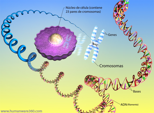
Something clear after the first findings about normal human genome, it's the fact this consists of 23 pairs of chromosomes, those inherited by mother andthose inherited by  father, but in total there are 24 pairs of chromosomes since  2 correspond to the sexual chromosomes X and the and, Y which harnessed on XX if you are female and XY if you are a male.
 
This is part of the working material of the genetic, which is responsible of researching  heritage and vulnerability of specific the genotypic traits and the genes associated with them. On the other hand, the new science called  Genomics sees genome as a whole, even their multiple interactions with environmental factors, resulting in perceptible all the individual organism. For this purpose it has methodological tools that allow you to study at a time, globally or in parallel, a single biological sample, thousands of genes or their products (Velázquez, 2004).


These gene sequences control most bodily functions and structures, such as the creation of organs, connection between  neurons in the nervous system, skin color, color of eyes, etc.,  however, in order to those genes exert their specific action is required in addition to their structural and functional integrity the presence of a suitable environment. 
Other two fundamental genetic concepts are genotype and phenotype, the first refers to an individual's genetic constitution is  genome specific to an individual, in the form of DNA, while phenotype refers to what is related with  apparent characteristics as  factions,  color of eyes,  timbre of the voice. Then we can say,  genotype can be defined as the set of genes of an organism and the phenotype as a set of traits of an organism.

So the generation of  diversity of races and physical features is  made by a genetic recombination which undergoes each generation, but each individual is genetically different from everyone else (except if you have an identical twin), since the variety of ovules  or sperm that are formed along the life is so great, for practical purposes  only can say that none of them is equal to the other. Thus, mutations are the specific material of genetic diversity, but is is even greater and less controllable in species with sexual reproduction, which constantly faces  different genomes.

Genome (is a word made up of genes and chromosome) and is the totality of genetic material of an individual that contains the information for the operation and the development of a new body, since the ovule  is fertilized by the sperm until the end of life (Kaessmann & Pääbo, 2002; Velázquez, 2004).
With all that  information emerges with its own light the Human genome project (HGP), which constitutes the greatest scientific adventure of human biology and the genetic map  known. Through this project now we know an important basis of the medicine of the future. 

The Human Genome Project is an international research  whose ultimate goal is a complete description of the human genome, through DNA sequencing. It is known that the human genome consists of 3 billion base pairs, which can be comparable to an encyclopedia composed of thousand volumes, each with a thousand pages and 3,000 letters per page. If the bases would be the letters; the genes are the words and phrases,  while the chromosomes would be different volumes of the encyclopedia. Once completed the sequencing of the human mitochondrial genome, the next step was   investigating the nuclear genome. Given the scale of the effort that goes into the project, it represents what this is the first project of great science in biology (Velázquez, 2004).

But all this sequence is not easy, since the term human genome is used to describe all of the genetic information (DNA content) of human cells. In fact, encompasses two genomes: a complex, nuclear genome, and a simple, mitochondrial genome.

Research shows that nuclear genome  contains more than 99% of cellular DNA, has a total of 3000 Megabases (Mb) which are distributed among 46 chromosomes, 22 different autosomal pairs and two sex chromosomes, which can be distinguished through the application of chromosome banding techniques.

The number of genes containing the nuclear genome is estimated in a range that ranges between 30,000 and 150,000. While the human mitochondrial genome is defined by a single type of DNA. Its nucleotide sequence has already been fully established and consists of 16,569 base pairs in length that contain 37 genes. Unlike its nuclear counterpart, the human mitochondrial genome is extremely compact, approximately 93% of their DNA sequence is coding.

 In the nuclear genome, the percentage of coding DNA is only 3%, the remaining 97%, whose importance is a cause for controversy, it has been improperly called Junk  DNA. But, he found, however, that plays a key role in the normal function of the genome, the repair and regulation, and perhaps even in the evolution of multicellular organisms. On the other hand, public and private human genome project drafts have revealed that all human are identical in 99.8% (Glusman, Sosinky, Ben-Asher, Avidan, Sonkin and Bahar, 2001). 

But the effort to decode the human genome, has been an adventure that has been possible at different stages. The first began in the 1950s, when James Watson and Francis Crick in 1953 discovered the helical structure of DNA.

 Later multiple researchers are added with discoveries key as the Paul Berg and collaborators, who in 1972 created the first recombinant DNA molecule, and scientists from Harvard University and the United Kingdom developed a technique for sequencing DNA.

 In the Decade of the 80s in a joint effort between the universities of Stanford, Utah, Japan and other countries proposes a method for mapping the entire human genome. A genetic map consists of several genetic markers located nearby, one of whose order has been able to determine along each of the chromosomes, as a physical map, which becomes an enormous collection of small chromosome fragments also sorted according to its relative position in their corresponding chromosomes. 

After that, efforts focused on which markers and in which order they are in each one of the fragments of the physical map, similar as pieces of a puzzle, if two fragments share one or more genetic markers, this indicates that the two fragments are contiguous. Thus the genetic map to know the order that saved between if the fragments of the physical map (Velázquez, 2004).

With the development in 1985 of PCR (polymerase chain reaction) by Kary Mullis and his  partners to replicate DNA,  different studies were performed to sequence models of micro-organisms. But it was until 1995 that was published genetic mapping of the first living organism: Haemophilus influenzae which consisting of 1740 genes. This technique aims to obtain a large number of copies of a particular DNA fragment, on the basis of a minimum; in theory just starting from a single copy of this original fragment,  it's possible to amplify a fragment of DNA.  Its utility is so huge because  after amplification, it is much easier to identify with a very high probability of disease-causing by bacteria or viruses, identify people (bodies), or do scientific research on the amplified DNA (Bartlett & Stirling, 2003).

References: 

Bartlett, D. & Stirling, H.  (2003) A Short History of the Polymerase Chain Reaction. Methods Mol Biol. 226:3-6.

Glusman, G. Sosinky, E., Ben-Asher, N. Avidan, D. Sonkin, A Bahar, D. (2001) Sequence, structure and evolution of a complete human olfatory receptor gene cluster. Genomics. 63: 227-245.

Kaessmann, H. & Pääbo, S. (2002) The genetical history of humans and the great apes. Journal of Internal Medicine. 251. 1-18.

National Human Genome Research Institute (SF) A brief history of the Human Genome Project. Available at: http://www.genome.gov/12011239
Velázquez, A. (2004) Lo que somos y el genoma humano: des-velando nuestra identidad. Ediciones científicas universitarias. UNAM. FCE.
Strathern, P. (1999) Crick, Watson y el ADN. Siglo Veintiuno Editores. España.

Watson, J. (2000) La doble hélice. Alianza Editorial. Madrid.

Senin, 24 Juni 2013

Genes and genetics, a bit of history

Everybody talks about genes these days, mainly keeping this old and crazy idea that genes can keep  hidden secrets, with no possibility of changes, but recent researches show how much we still have to learn from them, but first, a little bit of history...
 
Since far-off times humans have realized that many of their bodily and psychological characteristics as well as their diseases, concentrated in some families and that these traits tend to be inherited from one generation to another. Thus they figured out that genetics plays an important role in not only cognitive but human development in general. 
 
Learning, is not exempt from this influence, at one way or another, some skills are scheduled since before birth, but they will be modeled with  culture and environment influences that allows these primary skills. 

It's not the case beginning a debate on whether genetics has more weight than the environment or Vice Versa,  what this post wants to highlight is the existence of critical periods that can be, if necessary, be extended, thanks to brain plasticity and depend on learning strategies, ensure that a child can compensate the nature's shortcomings or caused by environment .

This is because sometimes is easy to put away the legacies that parents give their children genetically speaking, because it is controversial to talk about whether the role of the environment is more or less important than the genes (Velázquez, 2004). It is known that genes contribute much to the development, but the environment exacerbates or represses this input. To the end of the day, If how can we be  geniuses of music if we don't have access to a musical instrument?, why do certain activities are easier than others?, Why schools aren't able to create geniuses?. 

In order to answer all those questions, it would be interesting to explain a bit of history, to mention that studies on the genetic relationships began in the year of 1900 with some research from someone called Mendel, however is interesting to note that Mendel carried out his discoveries almost without a previous research history.

 
  The history of prior knowledge to Mendel studies dating back to biblical times, as in genesis, a reference that Jacob, employed a method is presented for their sheep and goats raised mottled offspring (Strathern, 1999), bt it was not until  1694, when Camerarius reported the existence of sexes in plants and carried out the first experiments on pollination.  


Years later, entre1761 and 1767 Kölreuter, had done  research on hereditary mechanisms using plants,  however, the findings of this research appeared to confirm the current genetic theories at the time (mixed bloods), since the intersection between different varieties of Nicotiana (Nicotiana tabacum is a herbaceous perennial plant, of the family of the Solanaceae, whose leaves occurs most of the tobacco consumed in the world today), originating a hybrid of intermediate appearance between the parents which concluded this was due to two factors that each parent contributed in the same proportion to the characteristics of the offspring. The factors found by Mendel is what we currently know as genes (Strathern, 1999) .


Between 1822 and 1824, three independent researchers, Knight, Goss and Seton, realized studies based on pea (peas), discovering the dominance of some characters in generation 1 and segregation of several hereditary features in generation 2; However, do not they studied later generations or the numerical distribution of the characteristics of each generation, so it was not possible to extend the data from their studies.
Returning with Mendel, it has been written that he was fond of plants and to improve crops, although there is evidence that initially worked with mice, activity that seemed somewhat out of place to their superiors, for what made a change of experimental material to over 14 species of plants (Mendel said in any writing that he did not believe that his superiors knew that plants have sex). 

The pea, plant that finally worked, is a hermaphroditic species which has no sex chromosomes and it is easy to cut the stamens, avoiding in this way the self fertilization. Traits studied in such plants by Mendel are stable, although not studied intermediate conditions. For the form of the seed employed 253 hybrid (filial generation 1, F1), which had been obtained from a cross between the smooth seed plants and plants of rough seed, which originated only smooth phenotype, and is inferred a single genetic formula (genotype, Ll). But when the hybrids are crossed, it obtained 7324 seeds, of which 5474 were smooth and rough skin 1850, which somewhat complicated first finds, this generation called it subsidiary 2, F2 (Morgado, 2001; Barahona, Suarez and Martínez, 2001).

However, despite all the essential knowledge to explain the genetic mechanisms, most of the information for your understanding was obtained in years after Mendel, with the discovery of the desoxyribonucleic acid (DNA) carried out by Johann Friedrich Miescher in 1868; nucleic acids name are due to Richard Altmann, who thus called them in 1879.

But in this historic journey, it's not possible to forget the findings of Watson and Crick, doctors who allowed to know that the genetic information is contained in the molecular structure of desoxyribonucleic acid (DNA) which is found on the inside of a nucleus of the cell, in structures called chromosomes. However their findings were made possible the work of Rosalind Franklin who was an expert in x-ray crystallography and that thanks to her work was possible to elucidate the structure of the DNA double helix. 

Thanks to the joint work of these researchers from Cambridge, is found that desoxyribonucleic acid is a type of macromolecule that is part of all living cells and that there is contained the genetic information necessary for the development and operation of known living organisms and some viruses, being responsible for its transmission of heritable traits to the next generation. Within the DNA is very organized and associated with different proteins, which forms the structure known as chromatin (Watson, 2000).

References: 

Barahona, A., Suárez, E. y Martínez, S. (2001) Filosofía e historia de la biología. México. Facultad de Ciencias. UNAM. 

Kaback, DB. (2013)  The modest beginning of one genome project. Genetics. 194 (2) 291-299.

Morgado, E. (2001) ¿Cuán Mendeliana es la patología genética humana?. Clínica y Ciencia vol. 1 Nº 3. 48-59.
 
Strathern, P. (1999) Crick, Watson y el ADN. Siglo Veintiuno Editores. España.

Vásquez  Laslop, M. y Velázquez Arellano, A. (2004) Genómica y el desarrollo de un nuevo individuo. En A. Velazquez (2004) Lo que somos y el genoma humano: des-velando nuestra identidad. Ediciones científicas universitarias. UNAM. FCE.
Watson, J. (2000) La doble hélice. Alianza Editorial. Madrid.

Sabtu, 08 Juni 2013

Study of the brain through images, cytoarchitecture and electrical activity

To continue our  journey around the annals of the history of the study of the brain, is worth mentioning that the neuroanatomy has undergone revolutionary changes in the last decades. That leap has been made possible thanks to the introduction of new imaging techniques such as: X-Ray computed tomography (CT, also called computed tomography CT), (PET) Positron Emission Tomography and Magnetic Resonance Imaging (MRI), thanks all these tools, it is possible to observe the structure and activity of the brain in unprecedented detail.

All those datas, specially volumetric and structural studies, CT and MRI are of crucial importance to understand brain differences and give answer to many questions, specially about neuro degenerative diseases (Allen, Bruss and Damasio, 2005). 

However, this neuro technological revolution did not begin from nothing or yesterday, all these amazing possibilities probably began  in 1783 with physician Luigi Galvani who was a passionate about anatomy,  and who had the idea of using electricity to move the leg of a dead frog. Does that sounds like Frankenstein?,  what was this idea of moving a leg of a dead frog?,  well, he began efforts to stimulate and visualize neural activity, and some explain this open a door to  analyze living brains now.

Many years later, in 1937, a neuroscientist Charles Sherrington could see points of light signals in neuronal activity, this surprised to a Spanish physiologist,  Jose Delgado, and  he used radio waves to study the brain of a bull in 1963.

But it was not until 1971 that voltage fluorescent studies begin to become popular, and during decade of  1980 with the fluorescent dye, it was possible to see how calcium concentration changes while it's synthesized in a cell, and this  opened doors for the study of the brain on a larger scale (Miesenbock, 2008).

I can't forget during  this tour, including another researcher that made important contributions to the study of the brain, so I must remember to Korbinian Broadman, who conducted research that allowed to distinguish 52 brain regions, thanks to his studies on cerebral cytoarchitecture made on histological samples that permitted find anatomical definitions of different brains, and his studies currently  are known as  areas of Broadman which are used to mapping the brain, since they have been associated with specific activities and brain functions  (Kandel, Schwartz & Jessel, 2000).

Among the researchers that devoted his time to understand the functions relate to anatomical  Broadmann's areas there is a name, Wilder Penfield, who was a Canadian neurosurgeon and during his surgeries he stimulated with an electric pulse small points on the surface of the brain at the same time he asked to patient if he or she  felt something (this was necessary to determine exactly which region he had to operate). 

He found out that when different regions of the brain are stimulated in this way, the patient could have different perceptions (Harrison, Ayling & Murphy, 2012). For example, when it was stimulated the occipital lobe, patient saw flashes of light, but if it was stimulated  the parietal area, persons could heard buzzing, or maybe noticed tingling in any part of the skin, or maybe if stimulation was done in another region the patient begin moving any part of the body. 

Based on these observations, Penfield made a neurocortex map,  since  he could find where each sensory modality was represented in a specific part of the cerebral cortex, and he figured out it was not only possible to relate  a cortical region for each sensory modality, but that each part of the body had assigned to a specific region in the cortex, but on the opposite side of the body; for example a patient responded to a  electrical  stimulation on the left motor cortex with a movement of right leg. 

Therefore all his research made  possible to recognize areas on the surface of the cerebral cortex and relate them to different processes, finding in each patient  areas  where it was possible to recognize a specific taste, a vivid childhood memory or the fragment of a long-forgotten melody (Sagan, 2003; Shreeve, 2005; Library of archives of Canada, 2009). 

One of the reported cases, is about a patient who during a brain surgery said, he could  listened with luxury of detail, a interpretation of a composition of orchestral when it stimulated an area specified in his brain with an electrode. Other patients experienced a specific emotion, a sense of familiarity or the full memory of an experience of childhood, all simultaneously, forgetting the fact they were in an operating room talking to the surgeon. 

Some patients explained these memories as small dreams, but did not appear in them the symbolism characteristic of  a reverie (Shepperd, 2004). In the specific case of electrical stimulation of the occipital lobe, which is related to the vision, a patient said to be seeing butterflies flying around, so real and palpable, that even lying on the operating table, stretched out the hand to catch them (Sagan, 2003). 

All this experiences gave a good idea how the brain is divided into areas and allowed to map and  understand much better those parcels of information processing.

 However, even though there have been isolated area and process specific, neuroscience still cannot understand how it is possible to carry out the processing of information and the storage and handling of data that day to allow us to understand the environment and adapt to it, and I think the main question of neuroscience is: how do electric and chemical impulses become subjective experiences?.

References:

Allen, J.; Bruss, j. & Damasio, H. (2005) structure of the human brain. Research and science. 23 - January. 68-75.

Harrison TC., Ayling OGS, Murphy, TH. (2012) Cortical Disctinct circuit mechanisms for complex forelimb movement motor and map topography. Neuron. 72 (2) 397-409.

Kandel, E.; J.H Schwartz, Jessell, t. (2000) h Principles of Neural Science. New York: McGraw-Hill.

Library Archives of Canada (2009) Famous Canadian Physicians. (Available online): http://www.collectionscanada.gc.ca/physicians/030002-2400-e.html.

Miesenbock, g. (2008) Lighting up the brain. Scientific American . Vol. 299. NUM. 4 34-43.

Sagan, C. (2003) the Dragons of Eden: speculations on the evolution of human intelligence. Barcelona. Criticism.

Shepherd, g. (2004) The synaptic organization of the brain. Oxford, University press.

Shreeve, j. (2005) Cornina complet brain: she is all... is here. National Geographic.  207  (3) 6-12.